Retinal imaging is a non-invasive diagnostic tool that captures a detailed view of the back of your eye. It’s a tool that is meant to supplement and greatly assist your traditional eye exam. Together with a prompt eye examination, you would benefit from retinal imaging immediately if you experience sudden flashes of light, new floaters, distorted vision, or if you have underlying conditions like diabetes. Identifying these signs early can prevent permanent vision loss.
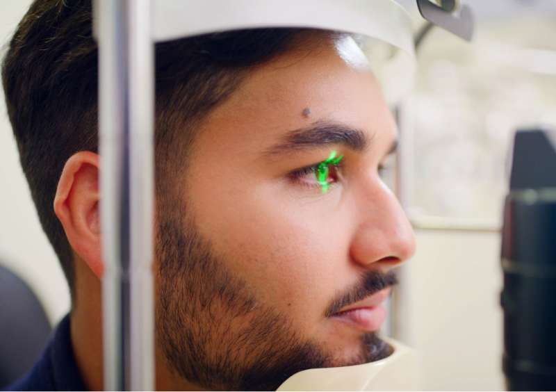
At Eyelike + Saigon Optical, we utilize state-of-the-art wide-field retinal imaging technology to detect sight-threatening conditions long before symptoms appear. Our advanced diagnostic approach allows our North York optometrists to capture a 200° view of your retina in a single, painless scan, providing a level of precision that traditional eye exams might miss.
What is Retinal Imaging and Why Is It Important?
Optomap Retinal imaging is a high-resolution digital screening that captures a 200° wide-field view of the back of your eye, including the retina, macula, optic nerve, and blood vessels. It is important because it allows optometrists to see deep structural details more in depth that may be hidden during a standard eye exam, enabling the early detection of life-altering diseases before vision loss occurs.
When Would You Benefit from Retinal Imaging?
If you’re wondering, "What Is a Symptom That Gives a Strong Indication of a Retinal Problem?”, you aren't alone. While many issues start silently, here are five critical signs that you may benefit from retinal imaging test during your eye exam:
1. Sudden Flashes of Light and New Floaters
You might see flickers of light like lightning streaks or a sudden shower of small dark spots. These are often symptoms of Posterior Vitreous Detachment (PVD), or in more severe cases, a retinal tear. Since the vitreous gel can pull on the retina and cause permanent damage, we use retinal imaging to scan the entire periphery of your eye to rule out sight-threatening emergencies due to detachments, tears, or breaks.
2. Blurred or Distorted Central Vision
Straight lines (like door frames or text on a page) appear wavy, or you have a "blank spot" in the center of your vision. Distortion in your central vision is a primary indicator of Age-Related Macular Degeneration (AMD) or macular edema. Because the macula is responsible for your sharp, detailed vision, any fluid buildup or thinning in this area requires immediate attention. Retinal imaging provides a high-resolution bird’s eye view of the macula to assist in detecting these microscopic changes before they become irreversible.
3. A "Curtain" or Shadow in Your Peripheral Vision
It feels like a dark shadow or a gray curtain is moving across your side vision. A loss of peripheral vision is a "red flag" for an escalating retinal detachment. Unlike a standard eye chart test, retinal imaging captures a wide-field view of the internal eye structure. This is the most effective way to identify if you are experiencing a retinal detachment and "closing in" on your field of view.
4. A Diagnosis of Diabetes or High Blood Pressure
You have been diagnosed with a systemic condition, even if your vision feels "normal." Systemic health issues like diabetes and hypertension directly affect the tiny blood vessels in your eyes.
Diabetic Retinopathy often has no symptoms in its early stages but can lead to sudden blindness. Retinal imaging acts as a preventative baseline, allowing your North York optometrist to monitor for leaking vessels or hemorrhages that indicate your condition is progressing.
